

Disease due to nontuberculous mycobacteria (NTM) is attracting a high level of interest,1 most likely because it is now recognized that NTM infection is increasingly frequent in both AIDS and non-AIDS populations.2 Lung disease is the most common clinical manifestation of NTM disease.3,4 Although the diagnostic criteria and treatment regimen for NTM disease have been the subject of several reports in the recent years,1,2,5 there is little information on the treatment responses of the patients in literature. We would like to report the results of our own series.
We retrospectively evaluated patients with pulmonary infections due to NTM who were treated with a therapy regimen from 2005 to 2011. Demographic data, predisposing factors, disease presentation and evolution were registered. All patients were clinically assessed and had sputum analyses performed monthly, and computed tomography (CT) of the chest was performed before and after treatment.
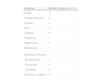
Eleven patients were included, 54.5% were male, and the mean age was 52 years (19–75 years). Four patients were smokers, five had previous pulmonary disease, four had gastroesophageal reflux, and two were frequent users of Jacuzzis. None of the patients were seropositive for HIV. The clinical and radiological presentations are shown in Table 1. The median time until diagnosis was 5 months.
The specimens used for microbiological diagnosis were sputum in 6 patients and bronchoalveolar lavage fluid in 5 patients.
The most frequently identified mycobacterium was M. avium complex (MAC) (54.5%). M. kansasii, M. peregrinum, M. scrofulaceum, M gordonae and M. fortuitum were identified in one patient each. Six patients were smear positive, of which five were submitted to nucleic acid amplification test (NAAT) for Mycobacterium tuberculosis; these tests were negative; apart from rare exceptions, a negative NAAT result for a smear positive specimen strongly indicates the presence of NTM species, excluding disease due to M. tuberculosis.6 Performing NAATs has a significant impact for smear positive patients because a negative NAAT result prevents patients from being subjected to unnecessary treatment for tuberculosis (TB) while waiting for culture results, which remain pivotal. Regarding the five patients for whom NAATs were performed, three waited for culture results, one started a broad therapeutic regimen for NTM and one was tested for NTM-specific sequences using polymerase chain reaction (PCR) technology. The one smear-positive patient who did not have NAAT was started on TB treatment until the culture results were available.
Nine patients were culture positive when NTM treatment was started, one was smear positive and positive for specific M. avium complex sequences by PCR, and the other started a broad therapeutic regimen which was adjusted when the culture results became available.
At the time at which the therapeutic regimen was started, all patients met the diagnosis criteria defined by ATS/IDSA.1 The therapy regimens were also chosen according to those guidelines, including the therapy for the patient with M. fortuitum. In vitro drug susceptibility tests were performed for this pathogen, and the treatment was chosen according to the results.
All patients were clinically assessed within 30 days, and clinical improvement was defined as the reduction or resolution of symptoms. Treatment was performed according to the guidelines – a treatment duration of 12 months starting from the first negative culture – except for one patient treated in 2005 who underwent only 8 months of therapy. Monthly sputum analyses were performed for the patients until microbiological conversion, which was defined as a negative sputum culture after treatment started. The time for culture conversion was the time between the first positive culture and the first negative culture.
Ten patients exhibited clinical improvement (symptoms reduction/resolution) within 3.6 months (1–13 months), the one patient who did not improve was also diagnosed with lung cancer. Mycobacterial conversion (negative sputum culture) occurred in all patients in 2.6 (0.5–7) months, and there was no evidence of bacteriological relapse. Seven patients exhibited radiological improvement that was detected after 6.6 months (4–12 months) of therapy, three patients exhibited no changes in the thorax CT scan, and one got worse.
Five patients have already completed the treatment, which lasted for an average of 14.2 months (8–18), and none of those patients have had any evidence of relapse (follow-up period of 13 months). One patient died due to vascular disease not related to the NTM infection and the other five are still under treatment.
We had a small number of patients who were treated for NTM, and the data for these patients should be integrated with the surveillance information regarding NTM which is limited in our country. In relation to the lack of HIV/immunodeficient patients, the small number of patients might be an important bias, but our results are consistent with those of a retrospective analysis performed in our geographical area7 regarding the NTM isolation in respiratory specimens and its clinical relevance; this previous study showed that although 20.6% of the patients with NTM isolations were HIV positive, the HIV status was not related to the NTM disease.
As reported in most series we found that MAC species were the most common species causing NTM lung disease. Male sex preponderance, mean age of disease presentation and the lack of symptoms specificity were also similar to previous reports.1 Clinical improvement was achieved within the expected time period (3–6 months).1 None of our patients had new positive sputum cultures after achieving sputum conversion. Interestingly, published studies on genotyping revealed that new positive sputum cultures for MAC after initial sputum conversion and culture negativity for at least 10 consecutive months while receiving a 3-drug, macrolide-containing regimen are more frequently due to re-infection than relapse.8
None of our patients had any drug intolerance or toxicity, even though they all underwent daily therapy. Nevertheless, it should always be emphasized that close monitoring is mandatory since treatment is often discontinued because of serious side effects.5
Please cite this article as: van Zeller M, et al. Micobactérias não tuberculosas – apresentação, diagnóstico e resposta ao tratamento. Rev Port Pneumol. 2010. http://dx.doi.org/10.1016/j.rppneu.2012.04.005.


