


A 67-year-old man presented to the Department of Neurointerventional Surgery with a 3-month history of left limb weakness. Chest computed tomography (CT) revealed an endobronchial lesion in the left lingular segmental bronchus (Fig. 1A–C). No respiratory symptoms, such as cough and sputum, were reported, although he had a smoking history of 50 pack-years. Fiberoptic bronchoscopy was performed, and the endobronchial mass measuring 1.5 cm × 1.0 cm × 0.8 cm in size was easily electro excised. Histopathological examination led to the diagnosis of mixed squamous cell and glandular papilloma (MSGP) (Fig. 1D) with the following immunohistochemical staining observations: epithelial cell CK5/6 (+) (Fig. 1E), CK7 (+), TTF-1 (+), NapsinA (−), basal cell P63 (+) (Fig. 1F), P40 (+), Ki-67 (1%, +). The patient was feeling well at the 2-month follow-up.
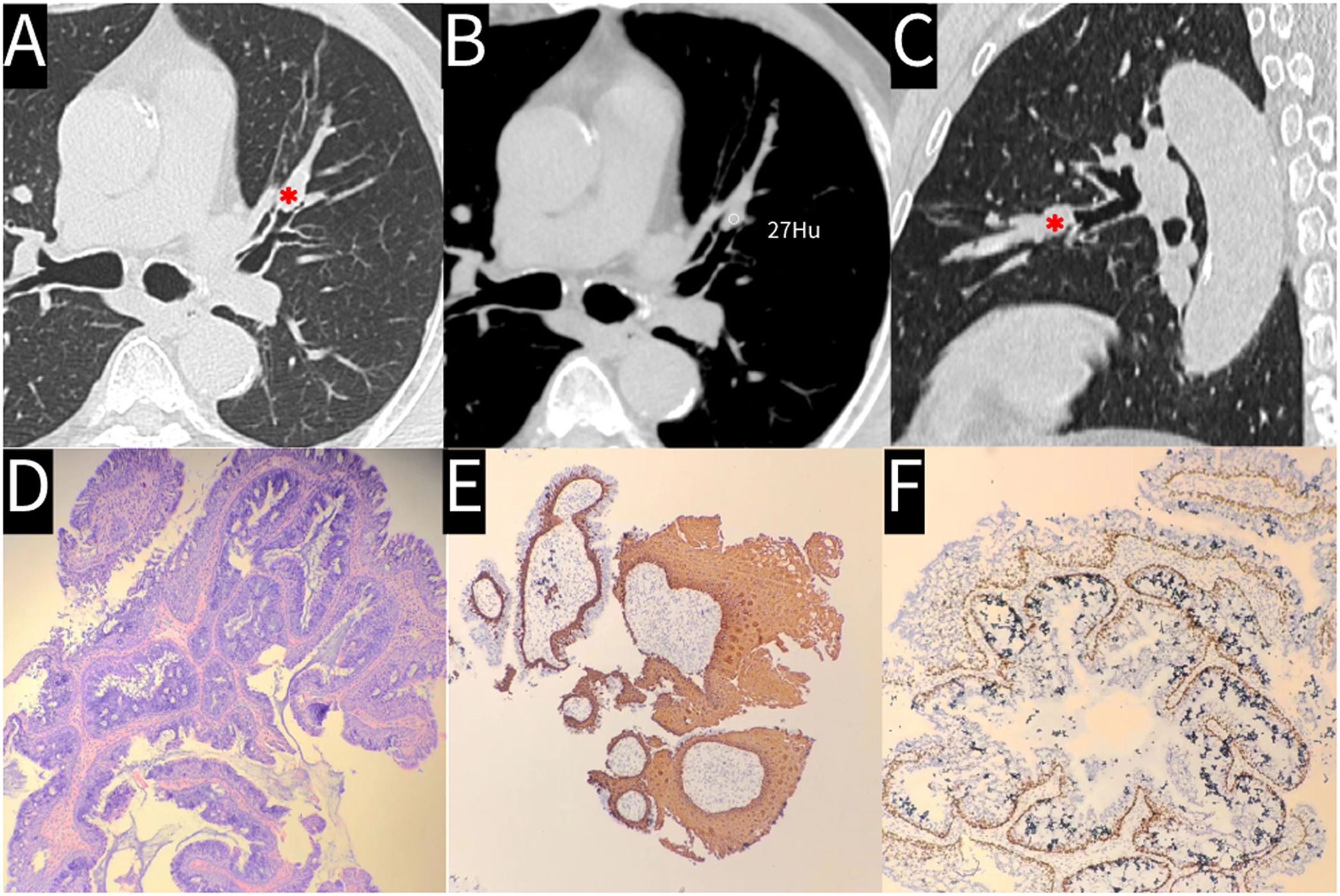
(A) Axial computed tomography (CT) with lung window showing an endobronchial mass in the left lingular segmental bronchus (asterisk). (B) CT value measurement of the endobronchial mass. (C) Sagittal CT with lung window showing the endobronchial mass (asterisk). (D) Hematoxylin and eosin-stained image showing branched fibrovascular cores in the center of tumor papilla lined by squamous and glandular epithelium. (E) Immunohistochemical staining showing CK5/6 expression in squamous and basal cells. (F) Immunohistochemical staining showing P63 expression in basal cells.
MSGP is a rare endobronchial papillary tumor characterized by a mixture of squamous and glandular epithelial cells. MSGP occurs more frequently in men who smoke and is commonly found in the left lingular segment.1 As the majority of MSGPs present with a lung mass on the CT image,2 endobronchial presentation of this MSGP is uncommon. Surgical resection is the mainstay of treatment as MSGP can potentially advance to carcinoma.2 In patients with limited endobronchial lesions, endoscopic therapy is an option.


