


The first step in the evaluation of patients with pleural effusion is to determine whether the effusion is a transudate or an exudate. An exudative effusion is diagnosed if the patient meets Light's criteria, although new formulas have been proposed.1
The serum to pleural fluid protein or albumin gradients may help improve categorization of the occasional transudate misidentified as an exudate by these criteria. If the patient has a transudative effusion, therapy should be directed toward the underlying cause like heart failure or cirrhosis. If the patient has an exudative effusion, attempts should be made to define the etiology. Pneumonia, cancer, tuberculosis, and pulmonary embolism account for most exudative effusions.
Cytomorphology is the hallmark for the diagnosis of malignancy.
The specificity of cytomorphology is very good and ranges from 91% to 100% with an average of 97%. Sensitivity ranges from 22% to 81% with an average of 58.2%.2
Morphologic distinction between reactive mesothelial cells and malignant cells can be difficult. Immunocytochemistry is a common adjunct method that serves to improve the sensitivity and specificity of cytology diagnosis.3 We used Ber-EP4 and calretinin in all malignant effusions. Ber-EP4 (epithelial cell adhesion molecule) is the most common marker expressed in all epithelial cells but not in mesothelial and hematopoietic cells.4 Calretinin is expressed in benign and malignant mesothelial cells but not in epithelial cells.
Five hundred malignant pleural effusion samples received for cytopathological examination from 500 patients hospitalized at the University Hospital of Heraklion Crete over a 5-year period were analyzed retrospectively.
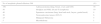
Table 1 shows distribution of malignancies in 500 neoplastic pleural effusions.
Distribution of malignancies in 500 pleural effusion specimens.
| No of neoplastic pleural effusions 500 | (%) | |
|---|---|---|
| 350 | Adenocarcinoma (lung, breast, ovary and GIT) | 70 |
| 50 | Hodgkin and NHL (B and T) lymphoma | 10 |
| 40 | Squamous carcinoma (lung, head and neck, larynx, genital track) | 8 |
| 30 | Neuroendocrine tumors (SCLC) | 6 |
| 25 | Malignant Melanoma | 5 |
| 5 | Mesothelioma | 1 |
Table 2 shows immunocytochemical markers expressed in studied malignancies.
Markers expressed in studied malignancies.
| Malignancy | Markers |
|---|---|
| Metastatic lung adenocarcinoma | CK7, TTF-1 |
| Metastatic breast carcinoma | CK19, Her2 neu, PR, ER receptors |
| Non-Hodgkin's lymphomas | PAX5 (PEL, B-cell type), CD4 (T-cell type) |
| Hodgkin's lymphomas | CD15, CD30 |
| Metastatic squamous carcinoma | Involucrin, CK5/6 |
| Metastatic neuroendocrine tumors | Chromogranin, Synaptophysin, NSE, CD56 |
| Malignant melanoma | HMB45, Melan-A |
| Malignant mesotheliomas | Calretinin, WT-1 |
Cytological examination of pleural fluids is often the first line of investigation to detect and type the neoplastic cells based on their subtle morphological features.5 Pleural metastases are more common in the visceral pleura and tend to be focal in the parietal pleura which is why pleural fluid cytology is a more sensitive diagnostic test than closed percutaneous pleural biopsy.6
The most common type of tumor to produce metastasis is the broad group of adenocarcinomas, most of them from lung, breast, ovary and GIT. In the present study the most frequent cause of malignant pleural effusion was lung. The neoplastic cells showed classical features of adenocarcinoma with large eccentric nuclei, nucleoli and vacuolated cytoplasm. These neoplastic cells expressed CK7 and TTF-1.
Metastatic breast cancer cells were often found in pleural effusions in our study expressed CK19, or Her2 neu and PR, ER receptors.
Pleural effusions were more commonly encountered in hematopoietic malignancies. In the present study NHL was the most common cause of pleural effusion due to lymphoma. The neoplastic cells expressed PAX5 in PEL and in all B-cell lymphomas and CD4 in T-cell lymphomas. In Hodgkin's lymphomas the neoplastic cells expressed CD15 and CD30 markers and the pathognomonic Reed-Stenberg cells were occasionally observed.
Among pleural fluids with metastatic squamous carcinoma SCC, the most common primary was lung, head and neck region, larynx and female genital tract. The presence of keratinized squamous cells and pearls were helpful diagnostic features. In this study the helpful markers for the diagnosis of SCC were involucrin and CK5/6.
The most common neuroendocrine tumor was the SCLC with isolated small cell or the characteristic Indian-file and molding of neoplastic cells and expressed Chromogranin, Synaptophysin, NSE and CD56.
Pleural effusions due to malignant melanoma were rare (5%). The malignant melanoma cells were isolated or composed of clusters with large nuclei and prominent nucleoli, and expressed HMB45 and Melan-A.
In 5 cases (1%) of primary malignant mesotheliomas the diagnosis was complicated because the malignant mesothelial cells resembled benign cells, but the clinical history of the patient was useful for the correct cytologic diagnosis. Bizarre enlargement, large nucleoli and abnormal cell forms, nuclear mitoses were found to favor malignancy. All malignant cells expressed calretinin and WT-1.
Genetic analyses of pleural effusion enhance the sensitivity for malignancy. Common features of early malignancy, which include DNA methylation, other mutations and microsatellite alterations, can be detected by polymerase chain reaction (PCR) and microarray techniques. Detection of epidermal growth factor receptor (EGFR) mutations in malignant cells can predict a favorable therapeutic response in patients with non-small cell lung cancer. On the other hand, recognition of Kristen ras (K-ras) oncogene mutations is a negative predictor of responsiveness to EGFR tyrosine kinase inhibitors.7
In 2007 anaplastic lymphoma kinase (ALK) rearrangement was discovered in approximately 5% of non small cell lung cancer (NSCLC), and ALK inhibitor (crizotinib) was rapidly approved in 5 years both in the USA and Japan. Today druggable oncogenes other than EGFR and ALK have been detected and development of specific inhibitors is underway.8
In conclusion, pleural effusion cytology with ancillary methods like ICC is a useful tool to detect malignant effusions and to suggest the type and the possible primary site of the tumor.
Conflicts of interestThe authors have no conflicts of interest to declare.


