

Non-invasive ventilation (NIV) undoubtedly brings benefits to the management of patients with acute respiratory failure. Although the number of indications has expanded rapidly due to increasing confidence in managing the technique, the optimal location for using NIV is still a matter of debate1,2. The vast majority of published evidence is based on studies conducted in highly controlled settings such as respiratory intermediate care units (RICU) and intensive care units (ICU). These outcomes may not always be representative of everyday practice since the strict criteria used to select patients may result in the exclusion of the more elderly and those with major comorbidities. However, as RICU/ICU beds are limited in most countries, use of NIV in respiratory wards may be an appropriate solution in selected cases, resulting in better resource utilization.
In Portugal, there is limited data available on the use of NIV outside the RICU/ICU.3
The purpose of this letter is to report on our experience with ward-based NIV in the treatment of acute and chronic exacerbated respiratory failure. We retrospectively analyzed demographic and clinical data of all patients who had had a NIV trial in our respiratory ward during a two-year period (2012–2013). Multivariate analysis was performed on age, sex, pH and PaCO2 before NIV, FEV1 and number of exacerbations in the previous year, to identify predictors of prolonged ventilation time (ventilatory support for more than 9 days) or NIV failure (need for endotracheal intubation or death). Elective admissions for setting up domiciliary NIV and patients treated with Continuous Positive Airway Pressure were excluded.
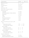
During the study period, 75 admissions were included, which corresponded to 65 patients, mostly male (54.7%) with a mean age of age 72±11.3 years. The demographic and clinical characteristics of all patients are summarized in Table 1. In the subgroup of chronic obstructive pulmonary disease (COPD) patients, the mean FEV1 was 42.7±16.3% of predicted, and 57% had had fewer than 2 exacerbations requiring hospitalization in the previous year.
Demographic and baseline clinical characteristics of the study group (n=75).
| Characteristics | Number±SD (or %) |
|---|---|
| Mean age (years) | 72±11.3 |
| Male sex | 41 (54.7%) |
| Smoking historya | 39 (52%) |
| LTOT | 47 (62.7%) |
| Domiciliary NIV | 13 (17.3%) |
| Hypercapnic respiratory failure | 68 (90.7%) |
| Indication for NIV | |
| AECOPD | 42 (56%) |
| COPD/Sleep apnea overlap syndrome | 12 (16%) |
| Pneumonia | 5 (6.7%) |
| OHS | 4 (5.3%) |
| ACPE | 3 (4%) |
| Neuromuscular disease | 3 (4%) |
| Ventilator weaning | 3 (4%) |
| Otherb | 3 (4%) |
| Mean arterial blood gases prior to NIV | |
| pH | |
| Hypercapnic patients | 7.29±0.10 |
| Non-hypercapnic patients | 7.43±0.05 |
| PaO2 (mmHg) | |
| Hypercapnic patients | 57±34 |
| Non-hypercapnic patients | 56±13 |
| PaCO2 (mmHg) | |
| Hypercapnic patients | 73±22 |
| Non-hypercapnic patients | 38±9 |
| Patients coming from | |
| Emergency department | 57 (76%) |
| Intensive/intermediate care unit | 18 (22.8%) |
ACPE, acute cardiogenic pulmonary edema; AECOPD, acute exacerbation of chronic obstructive pulmonary disease; COPD, chronic obstructive pulmonary disease; LTOT, long-term oxygen therapy; NIV, non-invasive ventilation; OHS, obesity hypoventilation syndrome.
At the time of admission, hypercapnic respiratory failure was predominant (90.7%), with a mean pH before NIV of 7.29±0.10. The most frequent indication for NIV was acute exacerbation of COPD (AECOPD, 56%), followed by exacerbation of COPD/sleep apnea overlap syndrome (16%), pneumonia (6.7%), obesity hypoventilation syndrome decompensation (5.3%), acute cardiogenic pulmonary edema (4%) and respiratory failure in neuromuscular disease (4%). In 4% of cases, NIV was used as part of ventilator weaning strategy.
The median time under NIV was 9 days (the cut-off used to define prolonged ventilation time) and the median hospital length of stay was 14 days. The ventilation time did not differ between groups according to previous domiciliary NIV use. In 14 NIV-trials (18.7%), adverse effects related to NIV were reported, most frequently nasal bridge ulcers and mucosal dryness. None of these complications caused procedure withdrawal. NIV failed in eight patients (10.7%): two were intubated and six died. Half of the failures were on NIV for AECOPD. The mortality rate in the study sample was 8%. The causes of death were pneumonia (n=4) and decompensation of comorbidities in patients with do-not intubate orders (n=2).
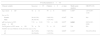
Among the variables studied, none was a predictor of prolonged ventilation time either in the global sample of patients or in the subgroup of AECOPD. The pretreatment pH was an independent predictive factor for NIV failure in the overall population (OR 0.92; CI 95% 0.85–0.99; p=0.034) (Table 2). We did not find any predictive factors for NIV failure in the AECOPD subgroup.
Predictive variables of NIV failure.
| Overall population (n=75) | |||||
|---|---|---|---|---|---|
| Clinical variable | Successn=67 | Failuren=8 | p value | Multivariate analysis | OR(95% CI) |
| Age, mean±SD | 72±11 | 79±8 | 0.052† | 0.165° | 1.07 (0.97–1.18) |
| Sex, n (%) | |||||
| Female | 29 (43.3%) | 5 (62.5%) | 0.302‡ | NA | NA |
| Male | 38 (56.7%) | 3 (37.5%) | |||
| pH level prior to NIV, mean±SD | 7.31±0.10 | 7.21±0.09 | 0.025† | 0.034° | 0.92 (0.85–0.99) |
| PaCO2prior to NIV, mean±SD | 70±24 | 71±15 | 0.810† | NA | NA |
| Number of exacerbations in the previous year, n (%) | |||||
| <2 | 48 (71.6%) | 7 (87.5%) | 0.092‡ | 0.152° | 5.8 (0.52–65.6) |
| ≥2 | 19 (28.4%) | 1 (12.5%) | |||
| AECOPD group (n=42) | |||||
|---|---|---|---|---|---|
| Clinical variable | Successn=38 | Failuren=4 | p value | Multivariate analysis | OR (95% CI) |
| Age, median (máx; mín) | 79 (92; 50) | 81 (88; 69) | 0.440¥ | NA | NA |
| Sex, n (%) | |||||
| Female | 16 (42.1%) | 1 (25%) | 0.507‡ | NA | NA |
| Male | 22 (57.9%) | 3 (75%) | |||
| pH level prior to NIV, mean±SD* | 7.29±0.08 | 7.22±0.09 | 0.214† | NA | NA |
| PaCO2prior to NIV, mean±SD* | 79±20 | 82±6 | 0.516† | NA | NA |
| Number of exacerbations in the previous year, n (%) | |||||
| <2 | 20 (52.6%) | 4 (100%) | 0.069‡ | NA | NA |
| ≥2 | 18 (47.4%) | 0 (0%) | |||
| Severity of airway obstruction | |||||
| Mild to moderate (FEV1 ≥50%) | 10 (26.3%) | 2 (50%) | 0.201‡ | NA | NA |
| Severe to very severe (FEV1 <50%) | 23 (60.5%) | 1 (25%) | |||
AECOPD, acute exacerbation of chronic obstructive pulmonary disease; FEV1, forced expiratory volume in 1 second; NA, not applicable; NIV, non-invasive ventilation.
Previous studies have shown that NIV delivered in the ward is effective in selected populations with reported success rates ranging between 66% and 85%.3–8 In a Portuguese study including patients with severe AECOPD treated in a respiratory ward, Oliveira et al. obtained a high success rate of NIV treatment (82%).3 Improvement in arterial blood gases in the first 24h was a determinant factor for NIV outcomes. On the other hand, Mclaughlin et al. have shown a lower success rate (66%) when using ward-based NIV with 392 AECOPD patients.8 However, these results may reflect differences in the severity of respiratory failure of the patients concerned (mean baseline pH of 7.24 versus 7.29 in our sample).
The importance of pretreatment pH level as a prognostic marker of NIV effectiveness in the ward was first highlighted by Plant et al., who found that patients whose pH was below 7.30 had significantly higher failure rates and in-hospital mortality.4 Our data also suggest that pretreatment pH is a useful predictor of NIV failure and should be considered when choosing the appropriate level of monitoring for each patient.
Other variables frequently reported as predictors of failure are pretreatment respiratory rate, Glasgow Coma Score, Acute Physiology and Chronic Health Evaluation (APACHE II), absence of early improvement in blood gases and excessive secretions.9,10 Given the retrospective nature of our study, the fact that severity scores for all patients were not available and the lack of standardized protocols guiding the use of NIV in our hospital, we were not able to evaluate the impact of those variables on the efficacy of NIV.
It is important to note that this is a single-center retrospective study, where NIV was delivered by experienced respiratory physicians and well-trained personnel. Thus, our results may not be reproducible in other general hospital wards.
We hope that the present study can fill a gap in Portuguese published data, giving a “real life” picture of the indications, outcomes, complications and clinical variables associated with failure of NIV in non-critical care setting.
In conclusion, our results suggest that NIV can be safely implemented in respiratory wards with a high success rate. It is our impression that successful implementation of NIV outside the RICU/ICU depends mostly on the monitoring capabilities of the ward and personnel resources available.
Conflicts of interestThe authors have no conflicts of interest to declare.


