


Bronchial artery embolisation (BAE) becomes a mainstay in the treatment of hemoptysis.
ObjectiveTo characterise patients with hemoptysis undergoing bronchial artery angiography (BAA) for embolisation, evaluating outcomes.
MethodsWe retrospectively evaluated patients with acute severe or chronic recurrent hemoptysis admitted to the Pulmonology department and submitted to BAA for purpose of embolisation.
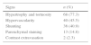
ResultsA total of 88 patients were submitted to BAA, 47 (53.4%) were male, with a mean age of 61.4±15.8 years. In 64 (72.7%) patients, hemoptysis presented as chronic recurrent episodes. Hemoptysis was considered severe in 40 (45.5%) patients. Bronchiectasis (other than cystic fibrosis) (n=35; 38.0%) and tuberculosis sequelae (n=31; 35.2) were the major aetiology for hemoptysis. The main angiographic abnormality was hypertrophy and tortuosity (n=68; 77.3%). BAE was performed in 67 (76.1%) of the 88 patients submitted to BAA. Immediate success was achieved in 66 (98.5%) patients. Recurrence of hemoptysis occurred in 25 (37.3%) patients, and was related to presence of shunting (p=0.049). The procedure-related complications were self-limited.
ConclusionOur results suggest that BAE is a safe and effective treatment for acute severe and chronic recurrent hemoptysis, supporting the current literature. Besides this, bleeding recurrence was relatively high, and correlated with presence of systemic pulmonary shunting.
Hemoptysis, defined as the expectoration of blood from the lung parenchyma or airways, is a common and alarming disease symptom.1,2 The circulation in the lungs—one of the richest in the human body—is provided via two separate vascular systems: the pulmonary and bronchial arteries.3 In cases of severe hemoptysis, 90% of the blood originates from the bronchial circulation, 5% from the pulmonary circulation and the remaining 5% from the non-bronchial systemic circulation.4,5 Hemoptysis is self-limiting and is approached effectively with conservative management in most cases.6 Nevertheless, it can be massive and life-threatening in 5–15% of cases,5 with a mortality rate of more than 50% in these cases if not treated appropriately.7 The currently available approaches for patients with hemoptysis are conservative medical treatment, bronchial artery embolisation (BAE) and surgery.8,9 The treatment of choice depends largely on the severity and urgency of the circumstances.
Bronchial artery angiography (BAA) with BAE is a minimally invasive procedure that has become a mainstay in the treatment of hemoptysis.10 It consists of selective bronchial artery catheterisation and angiography, followed by embolisation of any identified abnormal vessels to stop the bleeding. It has been used in patients with massive and recurrent hemoptysis,11 but rebleeding can emerge as a problem even after apparently adequate BAE.12,13
The purpose of this study was to identify and to characterise patients with hemoptysis who underwent BAA for embolisation in our centre and to evaluate their long-term outcomes.
Materials and methodsPatientsWe retrospectively evaluated 88 consecutive patients admitted due to hemoptysis to the pulmonology department of a university and to a tertiary care hospital, between January 2008 and December 2013. The patients all underwent BAA for embolisation purposes. The criterion for referral of these patients for BAE was an acute severe hemoptysis, as defined below, or a chronic recurrent hemoptysis requiring medical attention.
The information on blood volume expectorated by patients was limited; consequently, the assessment of severity was based on clinical criteria, including haematological compromise (a haemoglobin decrease of more than 1g), haemodynamic instability (defined as a systolic arterial pressure less than 90mmHg) and respiratory failure (defined as a PaO2 less than 60mmHg).14
After BAE, immediate success was defined as the absence of rebleeding during the first month after BAE. Recurrence was defined as hemoptysis occurring after BAE and requiring medical attention (unscheduled clinical visits, emergency room visits, admission or repeated BAE); recurrence was categorised according to timing in relation to the initial procedure in short-term (within 1 month) and long-term (more than 1 month) periods after BAE.
Bronchial artery embolisationTransfemoral bronchial arteriography was performed under local anaesthesia, percutaneously in the majority of the cases, using a 4F catheter. Both bronchial arteries and non-bronchial systemic arteries were opacified. The diagnostic angiographic injections were always selective into the bronchial, intercostal, subclavian, internal mammary, intercostobronchial and inferior phrenic arteries, using a microcatheter technique with a 2.7 F Terumo Progreat as the standard microcatheter.
Thoracic aortograms were performed mostly on those patients that did not undergo angio-CT prior to embolisation, in order to delineate the number, size and position of the bronchial arteries and to check for aberrant and ectopic bronchial arteries.
Several agents were used for embolisation: polyvinyl alcohol (PVA) particles, PVA hydrogel, microcoils and gelfoam, alone or in association, depending on angiographic abnormalities (hypertrophy and tortuosity of bronchial arteries, hypervascularity, parenchymal staining, contrast extravasation and arterioarterial or arteriovenous shunting) and the Interventional Radiologist's choice. PVA particles were by far the most commonly used embolic agents. Microcoils were used in association with PVA particles only in two patients, with hypertrophy, hypervascularity and shunting angiographic abnormalities.
Three Interventional Radiologists, two of them with more than 20 years of experience in bronchial artery embolisation and the other one with more than 10 years of experience in embolisation procedures did all the bronchial artery embolisation procedures.
Statistical analysisThe data were analysed using the statistical program IBM SPSS (Statistical Package for Social Sciences) version 21.0 for Windows. Differences between groups were analysed using the chi-square test or Fisher's exact test. A p value<0.05 was considered statistically significant.
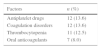
ResultsA total of 88 patients underwent BAA; 47 (53.4%) were male, the mean age was 61.4±15.8 years, and 38 (43.2%) patients had a smoking history. Apart from the underlying lung disease, additional risk factors for hemoptysis were identified (Table 1). Hemoptysis presented as chronic recurrent episodes in 64 (72.7%) patients and as a first episode in 24 (27.3%); it was considered severe in 40 (45.5%) patients.
Chest CTs (conventional or angiography) were available before BAA in 86 (97.7%) patients, and showed the possible causes of hemoptysis in 81 (94.2%). Eleven (12.5%) patients had thoracic CT angiography obtained less than one month before the procedure. Following initial stabilisation, bronchoscopy was performed in 53 (60.2%) patients: the bronchoscopy was flexible in 50 (94.3%), rigid in 1 (1.9%) and both flexible and rigid in 2 (3.8%). Bronchoscopy could identify the bleeding lobe in 20 (37.7%) patients, and endoscopic treatments to control bleeding were attempted in 3 (15%) of these patients, using cold saline instillation and topical adrenaline.
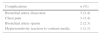
As shown in Table 2, bronchiectasis (other than that associated with cystic fibrosis) (n=35; 38.0%) was the major aetiology for hemoptysis. The aetiology could not be identified in 7 (8.0%) patients, even after BAA.
BAA was performed as an elective procedure in 72 (81.8%) patients and as an urgent or life-saving procedure in 16 (18.2%). During the procedure, the left bronchial territory was explored in 6 (6.8%) patients, the right bronchial territory in 19 (21.6%) and both territories in 63 (71.6%) patients. Non-bronchial systemic collateral arteries were systematically explored in 44 (50.0%) patients. The main angiographic abnormality was combined hypertrophy and tortuosity in 68 (77.3%) patients (Table 3). BAE was performed in 67 (76.1%) of the 88 patients who underwent BAA; BAE was performed in the left bronchial territory in 14 (20.9%) patients, right bronchial territory in 31 (46.3%) and both territories in 22 (32.8%). Several agents were used for embolisation: Polyvinyl Alcohol (PVA) particles (300–900 micron) in 57 (85.1%) patients, PVA hydrogel in 6 (9.0%), PVA particles and microcoils in 2 (3.0%), PVA particles and gelfoam in 1 (1.5%) and PVA hydrogel and Spongostan™ in 1 (1.5%). Immediate success was achieved in 66 (98.5%) patients. Despite this, recurrence occurred in 25 (37.3%) of these patients, with short-term recurrence in 5 (7.5%) and long-term recurrence in 20 (29.8%). The median time to recurrence was 161 days (1–955 days). Of all clinical and radiological features, shunting was the only factor favouring recurrence of hemoptysis (p=0.049). BAA was repeated in 16 (23.9%) patients in which recurrence occurred after initial BAE, and reembolisation was performed in 15 (93.8%) of those patients. The number of BAA procedures performed per patient during the period of the study ranged from 1 to 6. Pulmonary artery angiography as well as BAA was also performed in 6 (6.8%) patients, and 2 (33.3%) of these also underwent embolisation.
Procedure-related complications during the BAA were self-limited (Table 4). During the hospital stay, surgery was performed in 3 (3.4%) patients despite BAE (2 lobectomies and 1 bilobectomy), and 2 (2.3%) patients died (one of them due to fulminant bleeding). A total of 58 (86.6%) of the 67 patients who underwent BAE were eligible for follow-up after discharge, with a median follow-up period after the first BAE of 757 days (44–2185 days).
DiscussionIn our study, we found that BAE was a safe and highly effective procedure for hemoptysis control, with acceptable outcomes. These results are in accordance with those previously reported.15–18 Although the definition of immediate success of BAE varies in different studies, most reported success rates range from 80 to 90%.15 In our study, immediate success was achieved in 98.5% of our patients. Two recent retrospectives studies reported a success rate of 96.0% for immediate control of hemoptysis after BAE.19,20
Several definitions are used for hemoptysis severity, based on the amount of hemoptysis per 24-hour period. In our study, due to lack of data on the amount of bleeding, hemoptysis severity was based on the same criteria used in a previous study conducted at our centre.14 Using these criteria, almost half the patients in our study presented with severe hemoptysis, some of them warranting an urgent BAA and embolisation.
In agreement with Soares et al.,14 bronchiectasis and tuberculosis sequelae were the main associated pulmonary diseases in our study. Three other studies reported pulmonary tuberculosis (active and inactive) and bronchiectasis as the most common causes of life-threatening hemoptysis.21–23 Another study reported bronchiectasis and lung cancer (primary and metastatic) as the major etiologies of massive hemoptysis warranting BAE.15 Although lung cancer was the third commonest cause of hemoptysis in a previous study,14 only 2 (2.3%) of the patients in the present study who underwent BAA for embolisation purposes had this diagnosis. Mal et al. demonstrated that cancer-related hemoptysis had the highest failure rate and the worst long-term results after BAE.24 These findings and other data suggest that, in cancer-related hemoptysis, BAE may play a role as a temporary measure prior to surgical resection. More recently, Fugita et al. reported good results for BAA as a palliative measure in patients with advanced non-small cell lung cancer.25
During BAA, in the absence of an identified bleeding site, several sensitive findings can localise the bleeding source; these include vascular hypertrophy and tortuosity, neovascularity, hypervascularity, aneurysm formation and shunting (bronchial artery to pulmonary vein or bronchial artery to pulmonary artery). In our study, hypertrophy and tortuosity, hypervascularity and shunting were the main angiographic findings. Lee et al. reported similar results.26
Recurrence of hemoptysis after successful BAE is a common problem. The causes include recanalisation of embolised vessels, collateral circulation that feeds the bleeding lesion and underlying disease progression.15 In our study, long-term follow-up revealed a relatively high bleeding recurrence (29.8%). Inconsistent rebleeding rates have been reported in different studies. One study reported rebleeding in 28.0% of cases after successful BAE.27 Fruchter et al. reported bleeding recurrence in 57.7% of patients after successful BAE.15 The recurrence rates have been postulated to have a relationship with the aetiology of hemoptysis. Lung cancer, tuberculosis, aspergillosis and idiopathic bronchiectasis are associated with a high risk of bleeding recurrence.15,28,29 In these cases, BAE should be considered as a temporary and adjuvant therapy to surgery or as a specific medical therapy.
In our study, no significant statistical relationship was found between the aetiology and recurrence. In cases of early recurrence following an apparently successful BAE, systemic and pulmonary arterial contribution should be investigated.10,30 The benefit of performing a thorough investigation and embolisation of non-bronchial systemic collaterals at initial presentation has been confirmed, as their presence has a potential impact on recurrence of hemoptysis.10 Non-bronchial systemic collaterals were systematically sought in 44 (50.0%) of our patients.
It has also been suggested that characteristics of embolic agents affect the outcome of BAE. A variety of embolic materials, such as PVA, coils, gelfoams and microspheres, have been used for selective bronchial and non-bronchial systemic arterial embolisation in patients with hemoptysis. Each material has its own particular characteristics, with advantages and disadvantages. In our study, PVA was by far the most widely used embolic agent, so we were unable to perform a comparison between different embolic agents.
The complication rate for BAA and BAE has diminished gradually over the years, as a result of technical (super-selective technique) improvements and more appropriate embolic materials. The major complications include transverse myelitis, bronchial infarction, esophagobronchial fistula, ischaemic colitis, transient cortical blindness and stroke.12 Of these complications, the most feared is anterior spinal cord ischaemia due to the inadvertent embolisation of a spinal artery. The incidence of spinal artery ischaemia from BAE is reported as between 1.4% and 6.5%.31 Procedure-related complications occurred in a minority of patients in our study, and were self-limited. Anterior spinal cord ischaemia was not recorded and all patients were managed conservatively.
Since the advent of BAE, surgery has been gradually abandoned and is now used mainly in emergency scenarios, but it is still indicated in certain circumstances. Currently, surgery is mainly reserved for technical failure cases of BAE, early or repeated recurrences of hemoptysis despite BAE, or in extreme situations where the amount of bleeding or the patient's cardiopulmonary status is deemed life-threatening and prohibits transfer to an interventional radiology suite or precludes any related delays in management.5 Only 6.8% of our patients underwent surgical treatment despite BAE, which in part confirms the effectiveness of this technique.
Our study has some limitations. The retrospective nature of the study is the main limitation. The severity of hemoptysis could not be classified according to the volume loss, which may interfere with interpretation of the results and may limit possible comparisons with other studies. Some patients were lost to follow-up.
In conclusion, bronchiectasis (other than that associated with cystic fibrosis) and tuberculosis sequelae were the major aetiology for hemoptysis. Our results suggest that BAE is a safe and effective treatment for acute severe and chronic recurrent hemoptysis, supporting the current literature. In addition, bleeding recurrence was relatively high and was correlated with presence of systemic pulmonary shunting.
Ethical disclosuresConfidentiality of dataThe authors declare that no patient data appear in this article.
Right to privacy and informed consentThe authors declare that no patient data appear in this article.
Protection of human and animal subjectsThe authors declare that the procedures followed were in accordance with the regulations of the relevant clinical research ethics committee and with those of the Code of Ethics of the World Medical Association (Declaration of Helsinki).
Conflicts of interestThe authors have no conflicts of interest to declare.


